 |
||||||||
|
|
||||||||
 |
 |
|||||||
THE TORONTO CENTRE FOR PHENOGENOMICS
MOUSE IMAGING CENTRE |
|
|
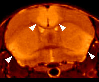
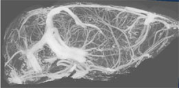
Cerebral Vascular DegenerationThere is growing concern about cerebral vascular disease and associated dementias given the aging population. Impaired cerebral blood flow is a feature of most senile dementias including Alzheimer's disease and vascular dementia. Identifying an animal model that reproduced important aspects of the impaired blood flow and blood vessel pathology would aid both in understanding these disorders and in developing effective treatments. It is of great interest, therefore, that we have obtained a new mouse model of progressive cerebral vascular disease that arose spontaneously in a mouse colony at the Jackson Laboratories [1]. These mice have been termed "Decrepit" or dcr because of their unhealthy appearance and shortened lifespan. T2w high resolution magnetic resonance images of dcr mutants show reproducible and bilaterally symmetric hyperintensities in areas of the brain consistent with the regions of multifocal necrosis identified using histological staining techniques (Figure 1). It is the goal of this research to explain the physical mechanism behind this observation and to describe the progression of disease in this mouse model. Our strategy is to examine the mice using high resolution 3D X-ray imaging (Figure 2), 2D arterial spin labeling (ASL) MRI and other specialized imaging techniques that have been adapted for mice. The dcr mutation has been mapped to mouse chromosome 9, syntenic to parts of human chromosome 3. The Jackson Laboratory will continue to try and identify the mutated gene responsible for the spontaneous appearance of this disease. Our work will lead to the definition of a new disease in mice. Our proposal is to establish whether this disease reproduces features of cerebral vascular disease seen in humans.
References1. Sproule TJ, Sled JG, Wentzell, J, Wang B, Henkelman RM, Roopenian DC, Burgess RW. A Mouse Model of Heritable Cerebrovascular Disease. PLoS One 5(12): e15327, 2010..
Research is funded by the Canadian Institutes of Health Research
|
||
|
© 2004 The Centre for Phenogenomics |
|