 |
||||||||
|
|
||||||||
 |
 |
|||||||
THE TORONTO CENTRE FOR PHENOGENOMICS
MOUSE IMAGING CENTRE |
|
|
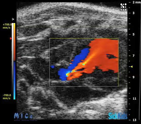
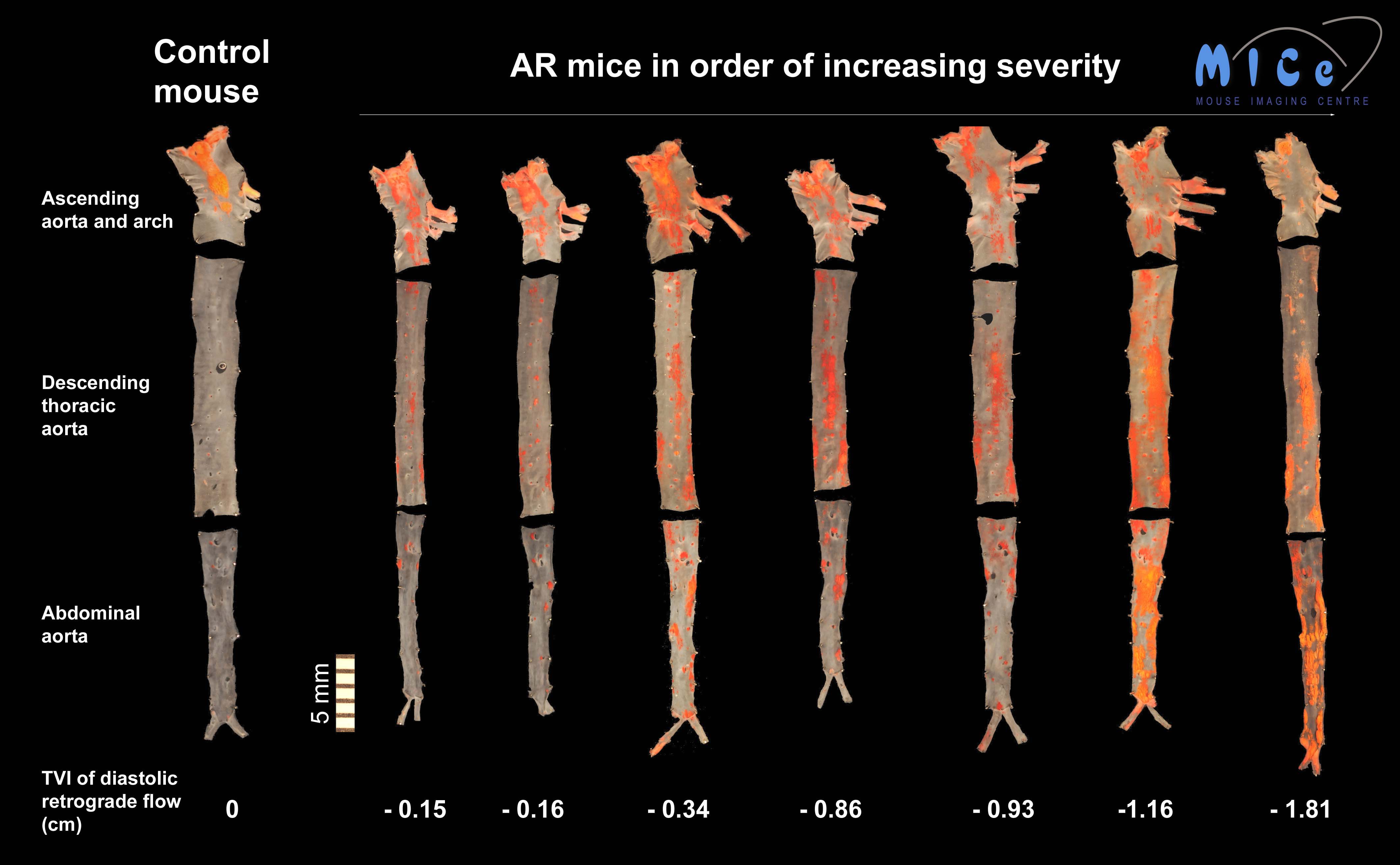
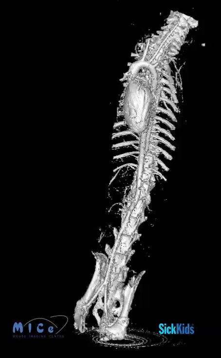
The Effect of Flow on Atherogenesis in MiceAtherosclerosis is one of the leading causes of death in Canada. This disease results in plaques in the vessel wall, which reduce and eventually block the blood supply to the heart and brain, causing myocardial infarction and stroke. The plaque lesions often occur along the inner side of curved vessels and lateral walls of vessel branches, probably due to the local blood flow patterns. However, the underlying mechanisms are not well understood. Discovering the relationship between the local blood flow pattern and the plaque formation will provide us better ideas for preventive and therapeutic strategies of atherosclerosis. We have developed a novel experimental model with altered blood flow patterns in the aorta by surgically damaging the aortic value to create aortic regurgitation (AR) (Figure 1) in a genetically-engineered mouse strain (ldlr-/-) which develops plaques when fed a high fat diet. As a consequence, the plaque distribution is dramatically changed throughout the entire aorta (Figure 2)[1,2]. In this project, we will use this model to evaluate the association between the blood flow pattern and the plaque formation throughout the aorta using the most advanced microimaging technologies including high frequency ultrasound, micro-CT (Figure 3), phase-contrast MRI and optical projection tomography (OPT), as well as the imaging-based computational fluid dynamic (CFD) simulations. At the same time, the effect of the altered blood flow pattern on the cellular and molecular events at the very early stage of atherosclerosis will be explored by fluorescently labeling specific biomarkers. In the end, we expect to find out the most crucial flow dynamic features responsible for the plaque initiation and its underlying mechanisms.
References1.Zhou YQ, Zhu SN, Foster FS, Cybulsky MI, Henkelman RM. Aortic regurgitation dramatically alters the distribution of atherosclerotic lesions and enhances atherogenesis in mice. Arterioscler Thromb Vasc Biol 2010;30;1181-1188. 2.Hoi Y, Zhou YQ, Zhang XL, Henkelman RM, Steinman DA. Correlation between local hemodynamics and lesion distribution in a novel aortic regurgitation murine model of atherosclerosis. Ann Biomed Eng. 2011 Jan 29. (Epub ahead of print) Research is funded by the Canadian Institutes of Health Research
|
||||||||||||
|
© 2004 The Centre for Phenogenomics |
|