 |
||||||||
|
|
||||||||
 |
 |
|||||||
THE TORONTO CENTRE FOR PHENOGENOMICS
MOUSE IMAGING CENTRE |
|
|
Manipulating Manganese-binding Proteins for Magnetic Resonance Imaging in the Mouse
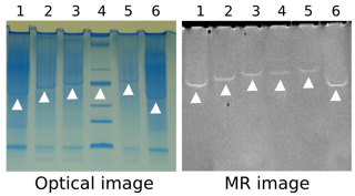
Manganese (Mn) is frequently used as a contrast agent for magnetic resonance imaging (MRI) studies in mice. Mn generates a distinct pattern of enhancement in the mouse brain that is useful for visualizing neuroanatomy. The persistence of the Mn contrast over time suggests that the pattern of enhancement is at least in part related to interaction between Mn and an as yet unidentified protein. We have developed a screen to identify proteins in the mouse brain that bind Mn in a way that generates signal enhancement on MRI. We are using in vitro studies to determine which of the identified candidates is most important in generating Mn contrast in the mouse brain. Frequently, to understand normal biology and disease, it is important to know when and where genes turn on and off (i.e., to map gene expression). This is often achieved by expression of fluorescent proteins in combination with light microscopy of fixed and sectioned specimens ex vivo. After identifying the most important Mn-binding proteins in the mouse brain, we intend to pursue the design of a Mn-based gene expression marker for MRI that can be used in vivo. Research is funded by the Natural Sciences and Engineering Research Council of Canada
|
||
|
© 2004 The Centre for Phenogenomics |
|