 |
||||||||
|
|
||||||||
 |
 |
|||||||
THE TORONTO CENTRE FOR PHENOGENOMICS
MOUSE IMAGING CENTRE |
|
|
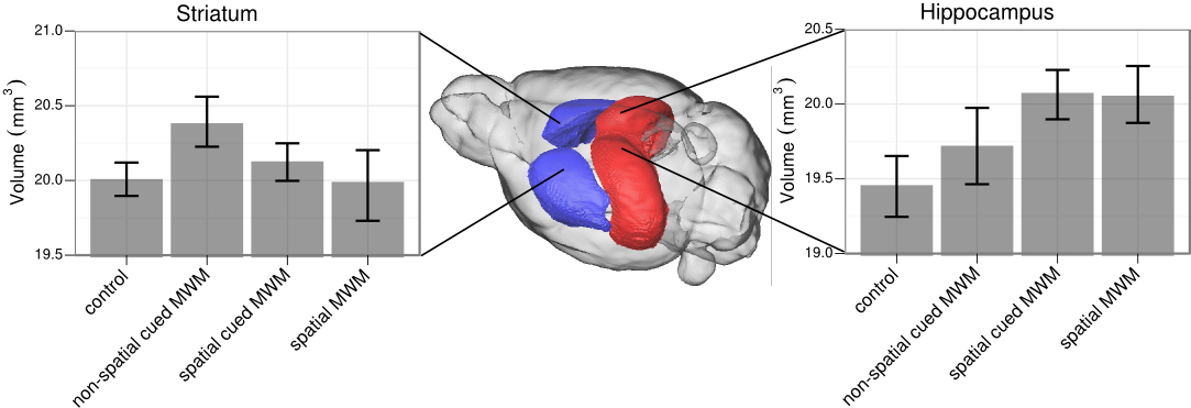
MRI of Learning and MemoryBrain imaging has made tremendous advances in identifying which areas of the brain are active during a given task. The longer term implications of learning and memory on the brain have, however, remained elusive. There are cellular changes that are known to occur in the learning process whose aggregate could produce a local volume displacement that can be captured with imaging. A recent study by our group set out to test for this phenomenon using Magnetic Resonance Imaging (MRI) in mice; we indeed were able to detect subtle brain volume changes when the mice learned to navigate a maze. The changes seen on MRI were cognitively specific, with the hippocampus growing in the spatial version of the maze and the striatum becoming enlarged in the non-spatial version, and colocalized with modulation of neuronal processes. Our goals now are to understand the cellular origins of how the brain changes with learning, to identify the best imaging and image processing techniques to capture brain plasticity, and to assess how aberrant plasticity is a factor in disease.
Research is funded by the Canadian Institutes of Health Research
|
||
|
© 2004 The Centre for Phenogenomics |
|