 |
||||||||
|
|
||||||||
 |
 |
|||||||
THE TORONTO CENTRE FOR PHENOGENOMICS
MOUSE IMAGING CENTRE |
|
|
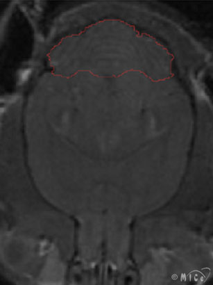
Image AnalysisAfter amassing so many images, the question is always: how do I analyze all this data? Most people require volumetric information about organs or substructures. The greyscale image does not yield such information immediately; the user must manually segment out the structure of interest. This can be done with either freeware (e.g. Display from the Montreal Neurological Institute) or commercially available software (e.g. Amira). We are currently developing automated image registration techniques to obviate the need for manual segmentation and to incorporate other methodologies to analyze the images, such as shape and principle components. Please see here for preliminary work in constructing a mouse brain atlas.
Other Links Montreal Neurological Institute, Brain Imaging Centre http://www.bic.mni.mcgill.ca
|
|||
|
© 2004 The Centre for Phenogenomics |
|