 |
||||||||
|
|
||||||||
 |
 |
|||||||
THE TORONTO CENTRE FOR PHENOGENOMICS
MOUSE IMAGING CENTRE |
|
|
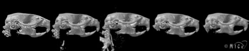
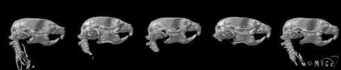
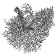
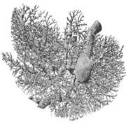
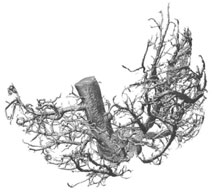
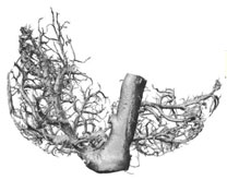
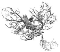
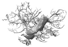
Micro-Computed Tomography (micro-CT)High-resolution X-ray computed tomography (micro-CT) produces detailed three-dimensional images of soft tissue and bone structure. In the case of soft tissue, the highest resolutions are achieved with the help of a contrast agent, which increases the X-ray attenuation of the tissue of interest. For this technique, the need for high X-ray doses to obtain the finest resolution precludes scanning of live specimens. At the Mouse Imaging Centre, a rotate-specimen type system supplied by General Electric Healthcare is used to examine excised organs and bone samples. This system is configured with a fixed X-ray source passing a cone of X-rays passing through the specimen and projecting onto a 2000 by 2000 element detector. As the specimen rotates, successive images are combined to build a 3D block of data showing the X-ray attenuation at each point within the specimen. Since images are created from the intact organ and the data generated is inherently 3D, this technology provides a powerful means of examining biological samples with complex structure. Developing computer tools to summarize the patterning of complex biological structures is an active area of research at the Centre. This technology facilitates comparison of specimens so as to identify anatomical differences due to disease or genetic manipulations.
References1. N.L. Ward, A.L. Haninec, P. Van Slyke, J.G. Sled, C. Sturk, R.M. Henkelman, I.R. Wanless, D.J. Dumont. Angiopoietin-1 Causes Reversible Degradation of the Portal Microcirculation in Mice: Implications for Treatment of Liver Disease. American J. Pathology 165(3):889-899, 2004. 2. J.G. Sled, M. Marxen, R.M. Henkelman. "Analysis of micro-vasculature in whole kidney specimens using micro-CT." 40th Annual Meeting of the SPIE, International Symposium on Optical Science and Technology, In Proceedings of the SPIE, vol. 5535, pp. 53-64, 2004.
|
|||||||||||||||
|
© 2004 The Centre for Phenogenomics |
|