 |
||||||||
 |
||||||||
|
|
||||||||
 |
 |
|||||||
THE TORONTO CENTRE FOR PHENOGENOMICS
MOUSE IMAGING CENTRE |
 |
|
|
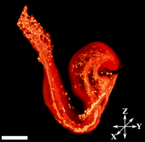
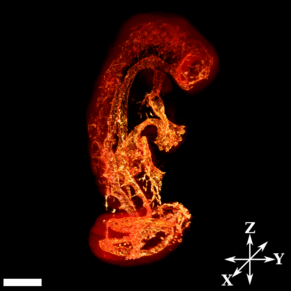
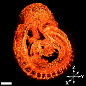
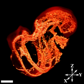
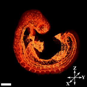
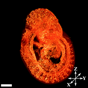
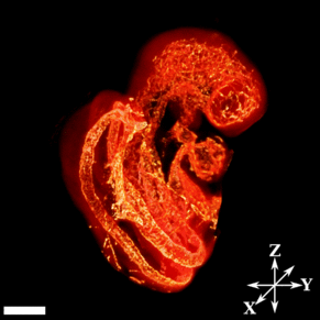
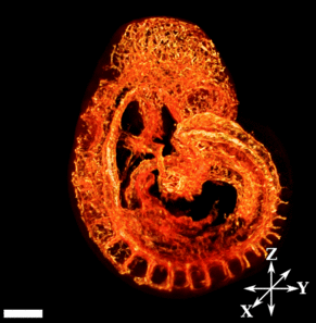
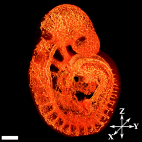
Vasculature Atlas of the Developing Mouse EmbryoA detailed description of this atlas can be found in:Walls JR, Coultas L, Rossant J, Henkelman RM. Three Dimensional Analysis of Vascular Development in the Mouse Embryo, PLoS ONE, e2853. (full text) Information and Instructions about the Developing Mouse Embryo Vasculature Atlas FilesThe vasculature atlas consists of Quicktime movies panning along three orthogonal axes through a 3-dimensional set created by merging the signals from the embryo autofluorescence and Cy3-PECAM (gray and hot metal, respectively). The X-Y plane corresponds roughly to the axial plane, the Y-Z plane to sagittal, and the X-Z to coronal. Overlaid on each movie is a simple surface render of the embryo with a solid green bar to denote the location of the current data slice through the 3D data set. The embryo age is denoted in somites, and all scale bars represent 200 microns. The image accompanying each data set consists of two overlapping volume renderings: the first is a red volume rendering of the embryo autofluorescence, and the other is a hot metal volume rendering of the embryo vasculature. All movies and images were generated with Amira 3.1 and the minc software tools. Software to view the the vasculature atlas movies is available for Windows, Mac, and Linux. The movies can be downloaded to your local disk using the Right-Click menu on PCs or Option-Click on Macs. Use of this material is granted provided the citation above is referenced and acknowledgements provided to the authors and the Mouse Imaging Centre.
Authors: Johnathon R. Walls, Leigh Coultas, Janet Rossant, R. Mark Henkelman
|
||||||||||||||||||||||||||||||||||||||||||||
|
© 2004 The Centre for Phenogenomics |
|
 Back to Mouse Atlas