 |
||||||||
|
|
||||||||
 |
 |
|||||||
THE TORONTO CENTRE FOR PHENOGENOMICS
MOUSE IMAGING CENTRE |
|
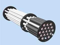
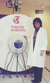
Magnetic Resonance Imaging (MRI)Magnetic resonance imaging (MRI) is a radiological tool used in the diagnosis of internal diseases in soft tissue, such as the brain and heart. In addition to excellent soft tissue contrast, the energy used in MRI is not harmful as found in X-ray. This makes this technology ideally suited for repeated studies in the same subject over a time course. In MRI, a magnetic field is used to align the protons or hydrogen nuclei found in water molecules in the subject. A radiofrequency pulse then excites the molecules, producing a signal that can be turned into images using a varying magnetic field, called a gradient. The same magnetic resonance principles can be applied for mice but since the mouse is so much smaller we need much larger fields to obtain the necessary high resolutions. Typical fields used are 4.7, 7, 9.4 and 11.7 T compared to clinical scanners at 1.5 T. At the Mouse Imaging Centre, we use a 7-T a Magnex Scientific magnet powered by a Varian Inc. Unity INOVA console. Even with the high field, it takes on the order of 10 hours to obtain specimen images of 60- µm isotropic resolution and 3 hours for live images between 110-µm to 160-µm isotropic resolution. To compensate for the long imaging times, we are developing a multi-mouse coil array that will image up to 16 mice at a time in our 30-cm bore (with gradients). Effectively, we will be reducing our imaging time to tens of minutes per mouse.
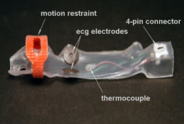
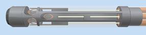
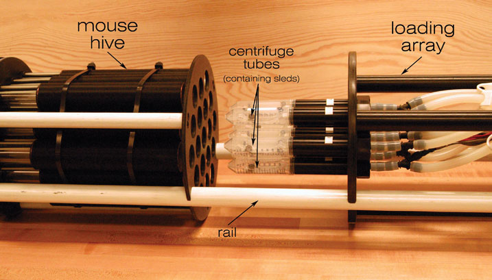
Techniques at MICeWhen performing biological experiments, what becomes immediately obvious is the large numbers of mice that are required in an experiment. It is common for studies to involve a few dozen mice. Given that it takes about three hours for a full 3D image to be taken of just the mouse head, a single experiment could take up an entire week. At MICe, we have developed a multiple-mouse imaging protocol (1) in which up to seven live mice are imaged simultaneously. We have capability to extend to sixteen mice in the future (Figure 1). To deal with the animal handling for the multiple mice, we have custom built a few pieces of equipment. In particular, we have built a large induction chamber to handle all mice simultaneously as well as a form-fitted “sled” (Figure 2). The preparation of 7 mice for imaging takes just under half an hour. To image multiple fixed organs, we have also built a three-coil solenoid coil array (Figure 3). Future developments include optimizing pulse sequences for multiple mouse imaging and cardiac imaging of multiple mice.
References1. Bock, N.A., Konyer, N.B. & Henkelman, R.M. Multiple-mouse MRI. Magn. Reson. Med. 49, 158-167 (2003). 2. Dazai, J., Bock, N.A., Nieman, B.J., Davidson, L.M., Henkelman, R.M. & Chen, X.J. Multiple mouse biological loading and monitoring system for MRI. Magn. Reson. Med. 52, 709-715 (2004). 3. Bock, N.A., Zadeh, G., Davidson, L.M., Qian, B., Sled, J.G., Guha, A. & Henkelman R.M. High-resolution longitudinal screening with magnetic resonance imaging in a murine brain cancer model. Neoplasia 5, 546-554 (2003). Other Links National Institutes of Health (NINDS) http://www.ninds.nih.gov/index.htm
|
|
|||||||||||||||||||||||

|
© 2004 The Centre for Phenogenomics |
|