 |
||||||||
|
|
||||||||
 |
 |
|||||||
THE TORONTO CENTRE FOR PHENOGENOMICS
MOUSE IMAGING CENTRE |
|
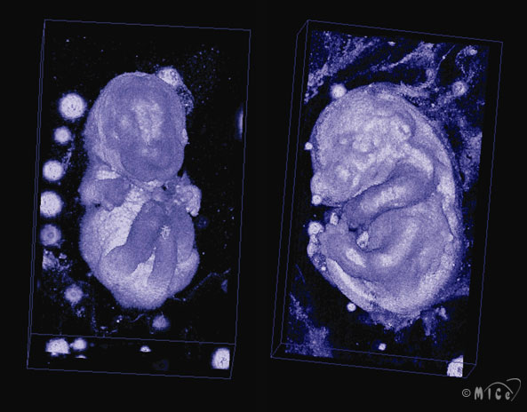
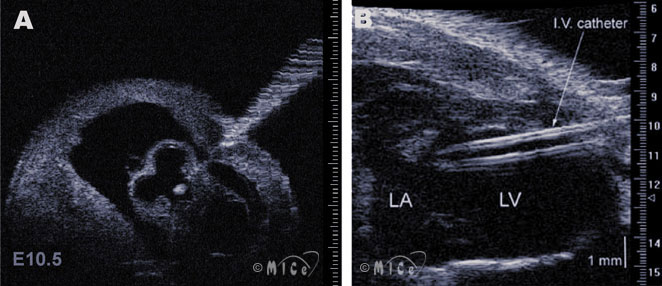
Ultrasound Biomicroscopy (UBM)Ultrasound biomicroscopy (UBM) is a new technology that uses very high frequency ultrasound (20~55 MHz or even higher, compared to 3~15 MHz in conventional clinical ultrasound systems) to noninvasively image small animals in biological research. At the Mouse Imaging Centre, we use a Vevo 660 UBM rolling cart developed by VisualSonics, Inc. The spatial resolution of a two-dimensional image is up to ~50 μm, with penetration depth of ~20 mm. The basic function modalities of UBM include:
Current applications of UBM in biological research:UBM provides a noninvasive approach for the morphological, functional and hemodynamic study on mice.
ReferencesFoster, F.S., Pavlin, C.J., Harasiewicz, K.A., Christopher, D.A., Turnbull, D.H. Advances in ultrasound biomicroscopy. Ultrasound Med. Biol. 26(1), 1-27 (2000). Zhou, Y.Q., Foster, F.S., Qu, D.W., Zhang, M., Harasiewicz, K.A., Adamson, S.L. Applications for multifrequency ultrasound biomicroscopy in mice from implantation to adulthood. Physiological Genomics 10(2), 113-126 (2002). Turnbull, D.H. Ultrasound backscatter microscopy of mouse embryos. Methods Mol. Biol. 135, 235-43 (2000). Zhou, Y.Q., Foster, F.S., Nieman, B.J. , Davidson, L. , Chen, X.J. , Henkelman, R.M. Comprehensive transthoracic cardiac imaging in mice using ultrasound biomicroscopy with anatomical confirmation by magnetic resonance imaging. Physiol. Genomics 18(2), 232-244 (2004). Zhou YQ, Davidson L, Henkelman RM, Nieman BJ, Foster FS, Yu LX, Chen XJ. Ultrasound-guided left-ventricular catheterization: a novel method of whole mouse perfusion for microimaging. Lab Invest 84(3):385-389 (2004). Other Links NYU Medical Center : http://www.med.nyu.edu/research/turnbd01.html
|
|
||||||||||

|
© 2004 The Centre for Phenogenomics |
|